


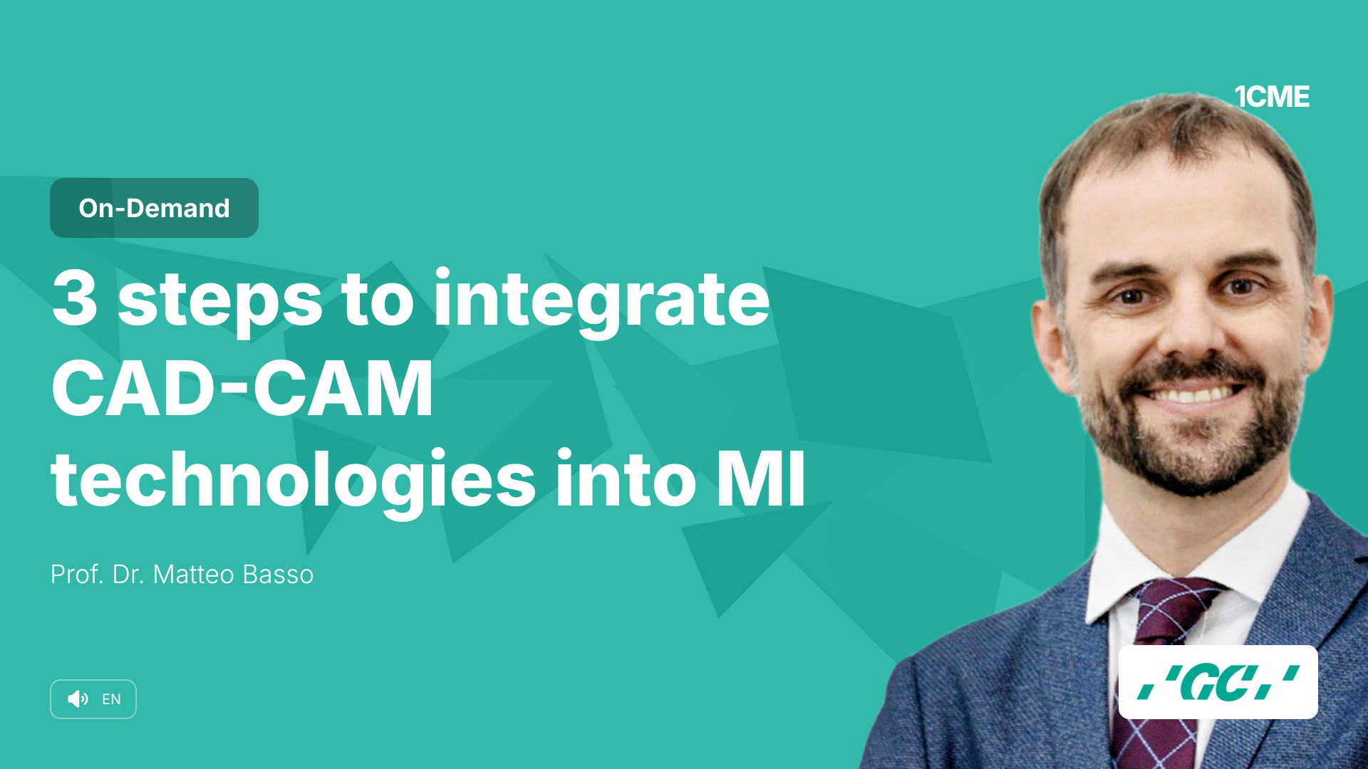
3 steps to integrate CAD-CAM technologies into MI
Highly predictable CAD-CAM devices are useful in fields such as conservative, preventive and MI dentistry. The digital information can be integrated to evaluate progression of pathologies, success of dental therapies, stability of clinical outcomes and evaluation of the impact of many risk factors.
The lecture will show which are the possible steps to easily get into a feasible integration for CAD-CAM technologies into Minimum Intervention, evaluating the costs-benefits aspects and several specific requirements of machinery. The use of an intraoral scanner is generally considered for crowns, bridges and implant dentistry. Another interesting field is the so-called “digital smile planning & design” where a 3D dental impression can be integrated with x-Rays images and digital photography for presenting a treatment plan and to pre-visualize the final result. But how many could be the integrations between the CAD-CAM technology and Minimum Intervention? The lecture include the following learning objectives:
- Learning Objective 1: Understand the requirements of a modern intraoral scanner, the ideal output 3D images and standards and definition of the importance of an open system for working in MI.
- Learning Objective 2: Evaluate the role of CAD-CAM systems in the diagnostic phase and the interpretation of data in determining the risk factors and the susceptibility of the patient to dental caries and gingival problems.
- Learning Objective 3: One of the most important aspect after setting preventive measures and therapies is the evaluation of their efficacy in preventive pathologies and the compliance of the patients in following our instructions. The lecture will provide several tools to make a safe check of our outcomes in the preventive phase.
- Learning Objective 4: Considering the same restorative treatment, it can be more or less successful depending on the patient’s susceptibility to pathologies and the existing dental structures. The lecture identifies several tips and tricks for using a digital scanner to check, rapidly and with few steps, the stability of dental restorations and periodontal procedures through 3D images.
Sprecher
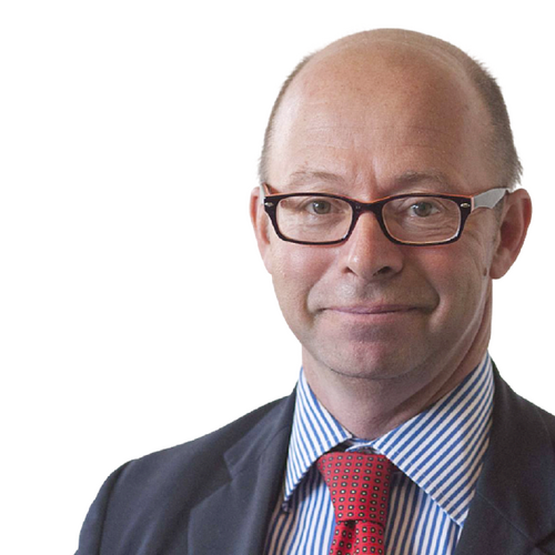
Prof. Dr. Matteo Basso DDS, PhD, MSc.
More courses with Prof. Dr. Matteo BassoGraduation with honor in Dentistry and Oral Prosthetic Rehabilitation at the “Università degli Studi” in Milan, Italy, in year 2000. In 2006 he gets PhD in “Innovative Techniques in Implant Dentistry and Oral Rehabilitation”. In year 2010: he obtains Specialty in Oral Surgery (MSc). In the period 2006-07 dr. Matteo Basso has been head of Conservative Dentistry Department at the University Dental Clinic of the Galeazzi Orthopaedic Institute in Milan. From January 2008 he became head of the innovative “Centre of Minimal Invasive, Aesthetic and Digital Oral Rehabilitation” (CROMED) in the same University, promoting new concepts and new digital approaches for prosthetics, dental caries management and tooth structures preservation. Actually Adjunct Professor of Dental Ergonomics and Business Management and Adjunct Professor of Periodontics for the modulus of Minimally Invasive Dentistry for the Graduation Course in Dentistry of the University of Milan. Active Member of ORCA (European Organisation for Caries Research), Active Member of TRAP-Group Italy (Tooth Respect and Prevention). Member and Founder of the international Minimum Intervention Advisory Board (MI-AB), Active Member of the Biodentistry.eu International group and Member of the IADR since 2007.